 CTICases
Granulomatosis with Polyangiitis (GPA) with Necrotizing Pulmonary and Skin Lesions
 ▶ Watch on YouTube — age-restricted, cannot embed here
[...]. Granulomatosis with Polyangiitis (GPA) with Necrotizing Pulmonary and Skin Lesions. CTICases. Published August 20, 2015. https://cticases.com/c/u9a9g
Imaging findings
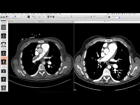
Chest radiographs showed large, rounded, non-segmental opacities in the lower lung zones. CT revealed extensive internal necrosis and cavitation within these large opacities, with a small right pleural effusion. The patient also had skin disease, with a lesion around the right elbow, and biopsy confirmed a granulomatous vasculitis. Elevated serum protease and ANCA positivity were present.
Key takeaways
Granulomatosis with Polyangiitis (GPA) is a systemic vasculitis characterized by necrotizing granulomatosis. Pulmonary manifestations include large, cavitating nodules with extensive internal necrosis. It often affects other organs like skin, kidneys, and sinuses. Elevated serum protease and ANCA positivity support the diagnosis.
AI-assisted summary — may contain errors. Verify against the source video. Learn more
More from this webinar
- Retained Wires from First-Generation DefibrillatorIatrogenic
- Hyperacute Pulmonary Infarction progressing to Acute PE with Pleural EffusionVascular
- Left Upper Lobe Anomalous Pulmonary Venous Stricture (Incidental)Congenital
- Lymphocytic Interstitial Pneumonia (LIP) in association with B-cell LymphomaNeoplastic
- Endobronchial Papillomatosis with Left Lower Lobe CollapseNeoplastic
- Esophageal Carcinoma with Radiographic CorrelationNeoplastic
- Cardiogenic Pulmonary Edema with Ground Glass OpacityVascular
- Hypersensitivity Pneumonitis (HP) in a Dog GroomerILD
See all cases from August 20, 2015 →
Related Autoimmune cases
- Fibrosing Mediastinitis (Sarcoidosis-related) Causing Pulmonary Vein Obstruction
- Microscopic Polyangiitis with Recurrent Diffuse Alveolar Hemorrhage
- Constrictive Bronchiolitis in Sjögren's Syndrome
- Nodular Sarcoidosis Mimicking Ground-Glass Opacity
- Progressive Fibrosing Sarcoidosis with Spontaneous Resolution of Mycetoma
- Nodular Sarcoidosis with Galaxy Sign