 CTICases
Aggressive Osteosarcoma with Pleural, Peritoneal, and Intravascular Metastases
 ▶ Watch on YouTube — age-restricted, cannot embed here
[...]. Aggressive Osteosarcoma with Pleural, Peritoneal, and Intravascular Metastases. CTICases. Published April 3, 2014. https://cticases.com/c/xha6u
Imaging findings
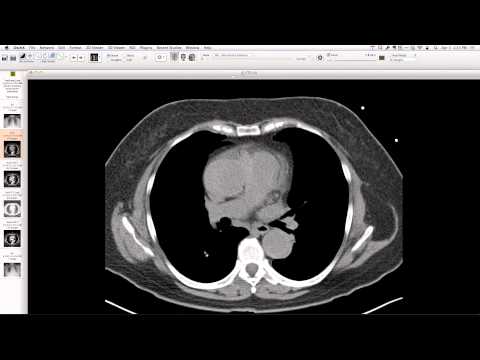
Diffuse, high-density, calcified, and ossified soft tissue nodules extensively involving both pleural spaces, fissures, and the peritoneum, with associated retroperitoneal lymphadenopathy. Intravascular metastases are also evident, with some nodules expanding pulmonary arteries. These findings represent progression from initial pleural metastases diagnosed in 2012, following a secondary osteosarcoma in a prior Ewing sarcoma radiation field.
Key takeaways
This case illustrates a highly aggressive osteosarcoma, particularly notable for its widespread metastatic pattern including peritoneal and intravascular dissemination in addition to extensive pleural involvement. The presence of intravascular metastases, seen as lesions expanding within pulmonary arteries, is a recognized but severe manifestation of osteosarcoma spread.
AI-assisted summary — may contain errors. Verify against the source video. Learn more
More from this webinar
- Williams Syndrome with Supravalvular Aortic Stenosis and Pulmonary Artery NarrowingCongenital
- Alveolar Capillary Dysplasia with Unroofed Coronary Sinus (Right-to-Left Shunt)Congenital
- Iatrogenic Esophageal Rupture from Blakemore Tube PlacementIatrogenic
- Primum Atrial Septal Defect (ASD)Congenital
- Pulmonary Alveolar MicrolithiasisMetabolic
- Esophageal Varices Anastomosing with an Inferior Pulmonary VeinVascular
- Unroofed Coronary Sinus (Isolated Finding)Congenital
- Complex Congenital Anomalies: Right Aortic Arch with Aberrant Innominate Artery, Total Anomalous Pulmonary Venous Return of Left Lung, and Cor Triatriatum SinisterCongenital
See all cases from April 3, 2014 →