 CTICases
Renal Cell Carcinoma with Pulmonary Artery Tumor Embolism
 ▶ Watch on YouTube — age-restricted, cannot embed here
[...]. Renal Cell Carcinoma with Pulmonary Artery Tumor Embolism. CTICases. Published March 4, 2016. https://cticases.com/c/ym9cw
Imaging findings
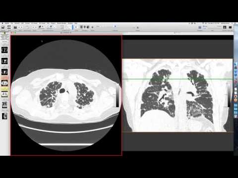
A patient with renal cell carcinoma had a very large tumor with extensive involvement of the IVC, crossing to the contralateral side. A CT of the chest showed a large lesion in the left lower lobe pulmonary artery, extending to a segmental vessel bifurcation. This was initially read as pulmonary embolism.
Key takeaways
Given the context of extensive renal cell carcinoma, the pulmonary artery lesion was concerning for tumor rather than glandular embolism. Frozen section confirmed tumor in the left pulmonary artery. The case highlights the importance of considering tumor embolism in patients with known extensive malignancy, even when a pulmonary embolus is suspected.
AI-assisted summary — may contain errors. Verify against the source video. Learn more
More from this webinar
- Fat Embolism SyndromeTrauma
- Dystrophic Calcification from Rhabdomyolysis and Pulmonary Interstitial EmphysemaMetabolic
- Nocardiosis in a Heart Transplant PatientInfection
- Pleuroparenchymal Fibroelastosis as a Manifestation of Graft-versus-Host DiseaseILD
- Synchronous Lung Cancer and Pulmonary HamartomaNeoplastic
- Variant Coronary Artery in the Ligament of MarshallCongenital
- Job's Syndrome (Hyper-IgE Syndrome) with Chronic Staphylococcal InfectionsAutoimmune
- Squamous Cell Carcinoma Obstructing Bronchus IntermediusNeoplastic
See all cases from March 4, 2016 →
Related Neoplastic cases
- Diffuse Large B-cell Lymphoma with Myocardial and Pulmonary Involvement
- Thymoma with Paraneoplastic Pure Red Cell Aplasia
- Hidden Lung Cancer
- Pulmonary Adenocarcinoma with Retained Acupuncture Needles
- Extensive Lymphoma with Extrapleural Paraspinal Involvement
- Obstructive Left Upper Lobe Collapse (Carcinoid and Adenocarcinoma)