 CTICases
Broncholithiasis
 ▶ Watch on YouTube — age-restricted, cannot embed here
[...]. Broncholithiasis. CTICases. Published July 17, 2014. https://cticases.com/c/9cxvt
Imaging findings
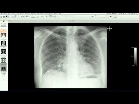
Chest CT shows a small, calcified broncholith within a left upper lobe bronchus associated with a surrounding ground-glass and solid nodular opacity, mimicking an adenocarcinoma. Ipsilateral calcified hilar and mediastinal lymph nodes are also present. Follow-up imaging reveals complete resolution of the ground-glass opacity, confirming it represented transient parenchymal hemorrhage.
Key takeaways
Broncholithiasis, typically caused by the erosion of a calcified lymph node into an adjacent bronchus (commonly from prior histoplasmosis or tuberculosis), can present with hemoptysis and cause localized parenchymal hemorrhage that closely mimics a neoplastic nodule or mass. Reviewing prior imaging for stable calcifications and obtaining short-term follow-up can confirm the transient nature of the surrounding hemorrhage.
AI-assisted summary — may contain errors. Verify against the source video. Learn more
More from this webinar
- Aortic Arch DissectionVascular
- Sclerosing PeriaortitisInflammatory
- Hypoplastic Left Heart Syndrome (Post-Norwood and Sano Shunt)Congenital
- Tetralogy of Fallot with Ductal Origin of the Left Pulmonary ArteryCongenital
- Migrating Pleural Mesothelial CystCongenital
- Chronic Diaphragmatic RuptureTrauma
- Bronchopulmonary DysplasiaCongenital
- Granulomatous-Lymphocytic Interstitial Lung DiseaseILD
See all cases from July 17, 2014 →
Related Other cases
- Spontaneous Pneumomediastinum and Pneumoretroperitoneum due to Macklin Phenomenon
- Esophagopericardial Fistula
- Benign Pneumatosis Intestinalis and Pneumoperitoneum in a Lung Transplant Recipient
- Costochondral Junction Ganglion Cyst
- Gastropericardial Fistula
- Incarcerated/Ischemic Morgagni Hernia (Recurrent)