 CTICases
Acute Type B Aortic Dissection with Unilateral Pulmonary Artery Hematoma
 ▶ Watch on YouTube — age-restricted, cannot embed here
[...]. Acute Type B Aortic Dissection with Unilateral Pulmonary Artery Hematoma. CTICases. Published January 14, 2016. https://cticases.com/c/afpws
Imaging findings
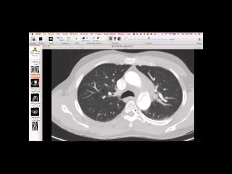
The initial radiograph showed a plump aortic arch, slight rightward tracheal deviation, and a fuller left pulmonary artery. CT revealed an acute type B dissection with a fenestration and mediastinal blood tracking along the left pulmonary arteries and left lung, accounting for the abnormal left hilum. There are peribronchial fluid cuffs of blood. The hematoma is conspicuously absent along the pulmonary veins. There is also complete collapse of the true lumen, leading to renal hypoperfusion and mesenteric ischemia, requiring a partial colectomy.
Key takeaways
This dissection likely involved an adventitial defect near the ductus remnant, explaining the unilateral hematoma tracking along the ligamentum arteriosum and pulmonary artery. The blood dissects within the connective tissue sheath that surrounds the pulmonary artery and companion bronchus, extending all the way to the small vessels in the lung. This is the first time observing unilateral blood tracking along the pulmonary arteries from a type B dissection. Normally, bilateral hematomas along pulmonary arteries are seen in type A dissections due to shared adventitial sheath near the root. Stenting the thoracic component of these dissections often opens the true lumen sufficiently to reperfuse the abdomen.
AI-assisted summary — may contain errors. Verify against the source video. Learn more
More from this webinar
- Fibrovascular Polyp of the EsophagusNeoplastic
- Ectopic Thymoma in the Lower NeckNeoplastic
- Pentalogy of CantrellCongenital
- Rapidly Progressive Interstitial Lung Disease in Amyopathic DermatomyositisILD
- Lymphangioleiomyomatosis (LAM) Mimicking Alpha-1 Antitrypsin DeficiencyILD
- Acute Severe Pulmonary Embolism Requiring Surgical EmbolectomyVascular
- Pulmonary Artery Pseudoaneurysm in Cystic FibrosisVascular
- Alpha-1 Antitrypsin Deficiency EmphysemaILD
See all cases from January 14, 2016 →