 CTICases
Fibrovascular Polyp of the Esophagus
 ▶ Watch on YouTube — age-restricted, cannot embed here
[...]. Fibrovascular Polyp of the Esophagus. CTICases. Published January 14, 2016. https://cticases.com/c/84p3c
Imaging findings
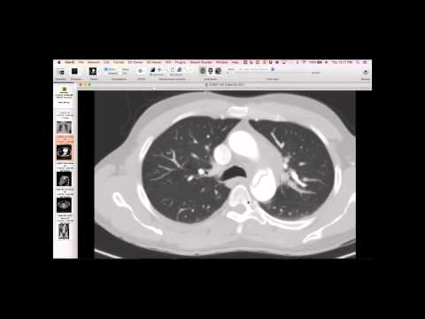
She has a mass in the region of her esophagus that looks like it's in the esophagus and there's areas that are fat. As you go further down there's more areas that are kind of mixed soft tissue. It expands the esophagus and extends from the thoracic inlet all the way down almost to the level of her left atrium, so it's a pretty elongated mass with different elements.
Key takeaways
She is an otherwise healthy 61-year-old nurse who had violent coughing up near asphyxiation. She noticed a large mass in the back of her throat and was able to forcefully reduce it back in her esophagus to regain her breathing capacity. This is a very rare tumor thought to arise from stromal elements in the cervical esophagus. Over the course of years, they elongate and continue to get larger, extending down the esophagus. A stalk is typically found near the cricopharyngeal region, which was found and allowed for removal.
AI-assisted summary — may contain errors. Verify against the source video. Learn more
More from this webinar
- Ectopic Thymoma in the Lower NeckNeoplastic
- Pentalogy of CantrellCongenital
- Acute Type B Aortic Dissection with Unilateral Pulmonary Artery HematomaVascular
- Rapidly Progressive Interstitial Lung Disease in Amyopathic DermatomyositisILD
- Lymphangioleiomyomatosis (LAM) Mimicking Alpha-1 Antitrypsin DeficiencyILD
- Acute Severe Pulmonary Embolism Requiring Surgical EmbolectomyVascular
- Pulmonary Artery Pseudoaneurysm in Cystic FibrosisVascular
- Alpha-1 Antitrypsin Deficiency EmphysemaILD
See all cases from January 14, 2016 →