 CTICases
Rapidly Progressive Interstitial Lung Disease in Amyopathic Dermatomyositis
 ▶ Watch on YouTube — age-restricted, cannot embed here
[...]. Rapidly Progressive Interstitial Lung Disease in Amyopathic Dermatomyositis. CTICases. Published January 14, 2016. https://cticases.com/c/wc6hs
Imaging findings
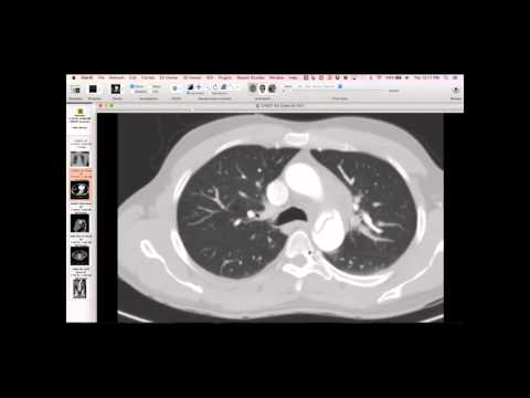
The patient presented with shortness of breath. Initial CT showed subtle lesions at the lung bases, including a particular blob that was biopsied and found to be non-infectious. A few weeks later, there was rapid progression with widespread ground glass opacities throughout the lungs. Intermediate CT scans showed findings suggesting organizing pneumonia, which can blend into NSIP.
Key takeaways
At autopsy, the lung disease was found to be an NSIP pattern. Serologic workup revealed anti-Ro 52 and anti-MDA5 antibodies, both associated with dermatomyositis. The patient did not have muscle findings, presenting with amyopathic dermatomyositis where interstitial lung disease was the leading symptom, along with eventual subtle skin findings. Anti-MDA5 antibody is associated with rapidly progressive amyopathic dermatomyositis, often with ILD leading the clinical picture. This presentation can include an acute lung injury pattern, often a combination of NSIP, acute lung injury, and organizing pneumonia.
AI-assisted summary — may contain errors. Verify against the source video. Learn more
More from this webinar
- Fibrovascular Polyp of the EsophagusNeoplastic
- Ectopic Thymoma in the Lower NeckNeoplastic
- Pentalogy of CantrellCongenital
- Acute Type B Aortic Dissection with Unilateral Pulmonary Artery HematomaVascular
- Lymphangioleiomyomatosis (LAM) Mimicking Alpha-1 Antitrypsin DeficiencyILD
- Acute Severe Pulmonary Embolism Requiring Surgical EmbolectomyVascular
- Pulmonary Artery Pseudoaneurysm in Cystic FibrosisVascular
- Alpha-1 Antitrypsin Deficiency EmphysemaILD
See all cases from January 14, 2016 →
Related ILD cases
- Lymphoid Interstitial Pneumonia (LIP) in Sjogren's Syndrome
- Acute Exacerbation of Idiopathic Pulmonary Fibrosis Triggered by Minor Trauma
- Common Variable Immunodeficiency with Lymphocytic Interstitial Pneumonia
- Interstitial Pneumonia with Autoimmune Features
- Desquamative Interstitial Pneumonia (DIP) in a Heavy Smoker
- Anti-Synthetase Syndrome (Anti-Jo-1) with Organizing Pneumonia