 CTICases
Pulmonary Artery Pseudoaneurysm in Cystic Fibrosis
 ▶ Watch on YouTube — age-restricted, cannot embed here
[...]. Pulmonary Artery Pseudoaneurysm in Cystic Fibrosis. CTICases. Published January 14, 2016. https://cticases.com/c/t79mg
Imaging findings
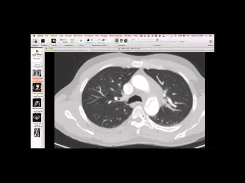
The patient with cystic fibrosis presented with hemoptysis. CT showed typical findings of CF and revealed a pseudoaneurysm arising from a pulmonary artery, which was opacified with contrast and surrounded by a hematoma within the lung. A central venous catheter was in place. Subsequent angiography and coiling confirmed and treated the pseudoaneurysm.
Key takeaways
This is a dramatic example of a pulmonary artery pseudoaneurysm in a patient with CF, which is less common than bronchial artery pathologies in this setting. While pseudoaneurysms are often attributed to mycotic causes in CF patients, this case highlights a rare presentation of significant hemoptysis. Typically, aneurysms and pseudoaneurysms are seen in conditions like Rasmussen's aneurysm or in the context of TB, rather than CF for a pulmonary artery pseudoaneurysm of this size.
AI-assisted summary — may contain errors. Verify against the source video. Learn more
More from this webinar
- Fibrovascular Polyp of the EsophagusNeoplastic
- Ectopic Thymoma in the Lower NeckNeoplastic
- Pentalogy of CantrellCongenital
- Acute Type B Aortic Dissection with Unilateral Pulmonary Artery HematomaVascular
- Rapidly Progressive Interstitial Lung Disease in Amyopathic DermatomyositisILD
- Lymphangioleiomyomatosis (LAM) Mimicking Alpha-1 Antitrypsin DeficiencyILD
- Acute Severe Pulmonary Embolism Requiring Surgical EmbolectomyVascular
- Alpha-1 Antitrypsin Deficiency EmphysemaILD
See all cases from January 14, 2016 →