 CTICases
Alpha-1 Antitrypsin Deficiency Emphysema
 ▶ Watch on YouTube — age-restricted, cannot embed here
[...]. Alpha-1 Antitrypsin Deficiency Emphysema. CTICases. Published January 14, 2016. https://cticases.com/c/ep3bh
Imaging findings
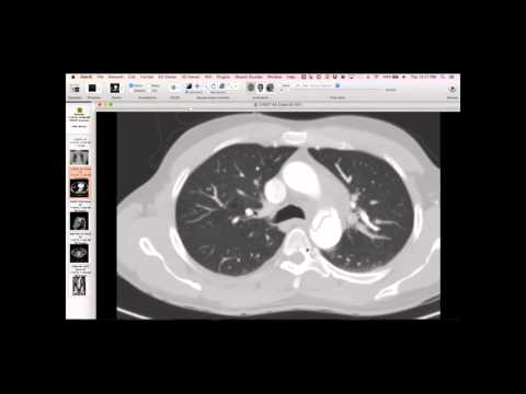
CT demonstrates confluent emphysematous holes, particularly in the left lower lobe, with very spindly vessels and large voids without intervening normal lung tissue. The characteristic centrilobular emphysema is also noted, but the panlobular destruction at the bases is most prominent.
Key takeaways
This case serves to contrast with LAM, where vessels in cystic areas may appear larger due to compressed normal tissue. In alpha-1 antitrypsin deficiency, the profound panlobular destruction leads to a more complete loss of capillary bed and spindly vessels, distinguishing it from LAM. The absence of normal intervening tissue and large voids are characteristic.
AI-assisted summary — may contain errors. Verify against the source video. Learn more
More from this webinar
- Fibrovascular Polyp of the EsophagusNeoplastic
- Ectopic Thymoma in the Lower NeckNeoplastic
- Pentalogy of CantrellCongenital
- Acute Type B Aortic Dissection with Unilateral Pulmonary Artery HematomaVascular
- Rapidly Progressive Interstitial Lung Disease in Amyopathic DermatomyositisILD
- Lymphangioleiomyomatosis (LAM) Mimicking Alpha-1 Antitrypsin DeficiencyILD
- Acute Severe Pulmonary Embolism Requiring Surgical EmbolectomyVascular
- Pulmonary Artery Pseudoaneurysm in Cystic FibrosisVascular
See all cases from January 14, 2016 →
Related ILD cases
- Lymphoid Interstitial Pneumonia (LIP) in Sjogren's Syndrome
- Acute Exacerbation of Idiopathic Pulmonary Fibrosis Triggered by Minor Trauma
- Common Variable Immunodeficiency with Lymphocytic Interstitial Pneumonia
- Interstitial Pneumonia with Autoimmune Features
- Desquamative Interstitial Pneumonia (DIP) in a Heavy Smoker
- Anti-Synthetase Syndrome (Anti-Jo-1) with Organizing Pneumonia