 CTICases
Acute Severe Pulmonary Embolism Requiring Surgical Embolectomy
 ▶ Watch on YouTube — age-restricted, cannot embed here
[...]. Acute Severe Pulmonary Embolism Requiring Surgical Embolectomy. CTICases. Published January 14, 2016. https://cticases.com/c/v9hbt
Imaging findings
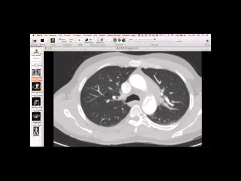
Radiograph showed marked enlargement of the heart, specifically the right atrium and ventricle, along with distension of the azygos vein and SVC, indicative of elevated CVP. Echocardiogram described a 'worm-like' mobile structure in the right atrium prolapsing through the tricuspid valve into the ventricle. A ventilation-perfusion scan showed numerous perfusion defects in both lungs, with only small areas of residual perfusion, indicating dramatic abnormality.
Key takeaways
This is a dramatic case of acute severe pulmonary embolism, confirmed by surgical embolectomy, which revealed emboli in the right atrium and both pulmonary arteries. Such severe PE can lead to pulmonary hypertension and significant cardiac strain. Surgical embolectomy is considered for severe cases, especially when there's an allergy to contrast preventing a CTPA, or when conservative management is insufficient.
AI-assisted summary — may contain errors. Verify against the source video. Learn more
More from this webinar
- Fibrovascular Polyp of the EsophagusNeoplastic
- Ectopic Thymoma in the Lower NeckNeoplastic
- Pentalogy of CantrellCongenital
- Acute Type B Aortic Dissection with Unilateral Pulmonary Artery HematomaVascular
- Rapidly Progressive Interstitial Lung Disease in Amyopathic DermatomyositisILD
- Lymphangioleiomyomatosis (LAM) Mimicking Alpha-1 Antitrypsin DeficiencyILD
- Pulmonary Artery Pseudoaneurysm in Cystic FibrosisVascular
- Alpha-1 Antitrypsin Deficiency EmphysemaILD
See all cases from January 14, 2016 →