 CTICases
Lymphangioleiomyomatosis (LAM) Mimicking Alpha-1 Antitrypsin Deficiency
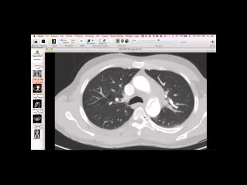 ▶ Watch on YouTube — age-restricted, cannot embed here
[...]. Lymphangioleiomyomatosis (LAM) Mimicking Alpha-1 Antitrypsin Deficiency. CTICases. Published January 14, 2016. https://cticases.com/c/mdzmh
Imaging findings
Chest radiograph and CT showed widespread cystic changes, initially suggestive of panlobular emphysema, particularly worse at the bases. However, closer inspection of the CT revealed relatively large, stretched vessels within the cystic areas, unlike the typically spindly vessels seen in severe alpha-1 antitrypsin deficiency emphysema. The cystic holes were also more homogeneous from top to bottom, lacking the clear apicobasal gradient seen in alpha-1 antitrypsin deficiency.
Key takeaways
This case highlights how LAM can mimic alpha-1 antitrypsin deficiency on initial imaging. A key distinguishing feature is the vascularity: in LAM, normal lung tissue is often interspersed between cysts, or compressed around them, maintaining some capillary bed, thus vessels may appear larger than expected for the amount of destroyed lung. In contrast, severe panlobular emphysema in alpha-1 antitrypsin deficiency typically shows very spindly vessels and confluent destruction with no normal lung tissue, giving a 'big void' appearance.
AI-assisted summary — may contain errors. Verify against the source video. Learn more
More from this webinar
- Fibrovascular Polyp of the EsophagusNeoplastic
- Ectopic Thymoma in the Lower NeckNeoplastic
- Pentalogy of CantrellCongenital
- Acute Type B Aortic Dissection with Unilateral Pulmonary Artery HematomaVascular
- Rapidly Progressive Interstitial Lung Disease in Amyopathic DermatomyositisILD
- Acute Severe Pulmonary Embolism Requiring Surgical EmbolectomyVascular
- Pulmonary Artery Pseudoaneurysm in Cystic FibrosisVascular
- Alpha-1 Antitrypsin Deficiency EmphysemaILD
See all cases from January 14, 2016 →
Related ILD cases
- Lymphoid Interstitial Pneumonia (LIP) in Sjogren's Syndrome
- Acute Exacerbation of Idiopathic Pulmonary Fibrosis Triggered by Minor Trauma
- Common Variable Immunodeficiency with Lymphocytic Interstitial Pneumonia
- Interstitial Pneumonia with Autoimmune Features
- Desquamative Interstitial Pneumonia (DIP) in a Heavy Smoker
- Anti-Synthetase Syndrome (Anti-Jo-1) with Organizing Pneumonia