 CTICases
Pleural Sarcoidosis
 ▶ Watch on YouTube — age-restricted, cannot embed here
[...]. Pleural Sarcoidosis. CTICases. Published July 17, 2014. https://cticases.com/c/ay888
Imaging findings
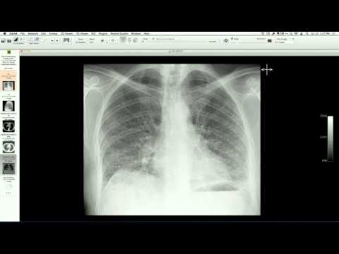
Chest CT demonstrates extensive, highly asymmetric nodularity and plaque-like thickening along the left visceral and parietal pleura (including the mediastinal pleural surface) associated with bilateral hilar, mediastinal, and subcarinal lymphadenopathy. Follow-up imaging after treatment shows near-complete resolution of the pleural pseudoplaques.
Key takeaways
Pleural involvement is a rare and unusual manifestation of sarcoidosis, occurring in less than 5% of patients, and typically presenting as pleural effusions or nodular pleural thickening (pseudoplaques) that can mimic pleural metastasis, lymphoma, or mesothelioma. Recognition of co-existing typical lymphadenopathy and classic biopsy findings (such as skin plaque biopsies) can prevent diagnostic delays and guide appropriate steroid or immunomodulatory therapy.
AI-assisted summary — may contain errors. Verify against the source video. Learn more
More from this webinar
- Aortic Arch DissectionVascular
- Sclerosing PeriaortitisInflammatory
- Hypoplastic Left Heart Syndrome (Post-Norwood and Sano Shunt)Congenital
- Tetralogy of Fallot with Ductal Origin of the Left Pulmonary ArteryCongenital
- Migrating Pleural Mesothelial CystCongenital
- Chronic Diaphragmatic RuptureTrauma
- Bronchopulmonary DysplasiaCongenital
- Granulomatous-Lymphocytic Interstitial Lung DiseaseILD
See all cases from July 17, 2014 →