 CTICases
Accelerated Complicated Silicosis
 ▶ Watch on YouTube — age-restricted, cannot embed here
[...]. Accelerated Complicated Silicosis. CTICases. Published January 14, 2016. https://cticases.com/c/hke4p
Imaging findings
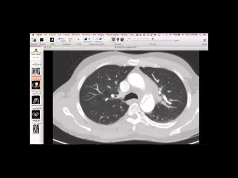
A 29-year-old stonecutter with only nine years of exposure presents with advanced silicosis. The radiograph shows complicated silicosis with large opacities on a background of small nodules and upper lobe volume loss. CT further demonstrates large opacities lacking internal bronchiograms, dense eggshell calcification in the lymph nodes, pseudoplaques along the pleural surfaces, and a background of small nodules predominantly in the upper zones but extending to the bases.
Key takeaways
This is an impressive example of accelerated complicated silicosis in a young individual after a relatively short exposure period. Despite the dramatic imaging findings, the patient was reportedly not severely symptomatic, possibly due to their young age and lung reserve. This presentation is less commonly seen in current practice.
AI-assisted summary — may contain errors. Verify against the source video. Learn more
More from this webinar
- Fibrovascular Polyp of the EsophagusNeoplastic
- Ectopic Thymoma in the Lower NeckNeoplastic
- Pentalogy of CantrellCongenital
- Acute Type B Aortic Dissection with Unilateral Pulmonary Artery HematomaVascular
- Rapidly Progressive Interstitial Lung Disease in Amyopathic DermatomyositisILD
- Lymphangioleiomyomatosis (LAM) Mimicking Alpha-1 Antitrypsin DeficiencyILD
- Acute Severe Pulmonary Embolism Requiring Surgical EmbolectomyVascular
- Pulmonary Artery Pseudoaneurysm in Cystic FibrosisVascular
See all cases from January 14, 2016 →
Related Exposure cases
- Silicone Injections and Pneumonitis
- Siderosis (Iron Deposition) from Occupational Exposure
- Necrotizing Fasciitis / Osteomyelitis from Heroin Skin Popping
- Pulmonary Langerhans Cell Histiocytosis (PLCH) - Classic Presentation
- Pulmonary Langerhans Cell Histiocytosis (PLCH) - Milder Spectrum (RBILD Transition)
- Bronchial Anthracofibrosis