 CTICases
Desquamative Interstitial Pneumonia (DIP) in a Heavy Smoker
 ▶ Watch on YouTube — age-restricted, cannot embed here
[...]. Desquamative Interstitial Pneumonia (DIP) in a Heavy Smoker. CTICases. Published October 22, 2015. https://cticases.com/c/jjbyj
Imaging findings
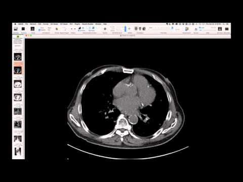
Chest radiograph shows a subtle diffuse fine reticular pattern. CT of the lungs reveals features of smoker's lung, including emphysema, with some larger cystic spaces potentially representing Langerhans cell histiocytosis. There is a diffuse background hyperattenuation throughout the lungs, most likely due to the accumulation of smoker's macrophages. Possible upper lung fibrosis is also noted.
Key takeaways
This case, in a heavy smoker, is a presumed diagnosis of Desquamative Interstitial Pneumonia (DIP), characterized by diffuse ground-glass hyperattenuation attributed to abundant smoker's macrophages, often co-existing with emphysema and possibly Langerhans cell histiocytosis. The findings represent a spectrum of smoking-related lung injury, and illustrate how these can combine to produce striking imaging.
AI-assisted summary — may contain errors. Verify against the source video. Learn more
More from this webinar
- Interrupted Aortic Arch, Type A with Aortopulmonary WindowCongenital
- Interrupted Aortic Arch, Type B Variant with Aberrant Subclavian ArteriesCongenital
- Post-Transplant Lymphoproliferative Disorder (PTLD), Polyclonal Polymorphic TypeNeoplastic
- Primary Effusion Lymphoma (PFL) in HIV-Positive PatientNeoplastic
- Apical Hypertrophic Cardiomyopathy with Apical AneurysmCongenital
- Double Saddle Pulmonary EmbolismVascular
- Primary Coccidioidomycosis with CavitationInfection
- Disseminated CoccidioidomycosisInfection
See all cases from October 22, 2015 →
Related ILD cases
- Anti-Synthetase Syndrome (Anti-Jo-1) with Organizing Pneumonia
- Fibrotic Organizing Pneumonia
- Restrictive Allograft Syndrome (Post-Transplant Upper Lobe Progressive Fibrosis)
- Bleomycin-Induced Severe Alveolar Damage with Barotrauma
- Hypersensitivity Pneumonitis (HP) in a Dog Groomer
- Methotrexate-induced Lung Toxicity (Organizing Pneumonia)