 CTICases
Metastatic Pulmonary Calcification
 ▶ Watch on YouTube — age-restricted, cannot embed here
[...]. Metastatic Pulmonary Calcification. CTICases. Published July 17, 2014. https://cticases.com/c/s5s24
Imaging findings
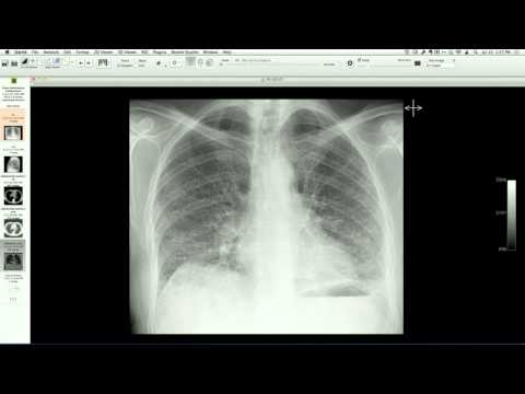
Dual-energy chest radiograph and high-resolution chest CT show upper-lobe-predominant centrilobular ground-glass nodules that spare the periphery of the secondary pulmonary lobules (asinar pattern). Bone scintigraphy demonstrates intense, diffuse uptake of the radiotracer within the upper lung zones, confirming the presence of metastatic calcium deposits in a patient with chronic renal failure.
Key takeaways
Metastatic pulmonary calcification is a systemic deposition disorder occurring in patients with chronic renal failure and secondary hyperparathyroidism. The CT findings of centrilobular ground-glass or air-space nodules can mimic infection, edema, or hemorrhage; however, intense tracer uptake on a bone scan is diagnostic and diagnostic of dystrophic/metastatic calcium deposition.
AI-assisted summary — may contain errors. Verify against the source video. Learn more
More from this webinar
- Aortic Arch DissectionVascular
- Sclerosing PeriaortitisInflammatory
- Hypoplastic Left Heart Syndrome (Post-Norwood and Sano Shunt)Congenital
- Tetralogy of Fallot with Ductal Origin of the Left Pulmonary ArteryCongenital
- Migrating Pleural Mesothelial CystCongenital
- Chronic Diaphragmatic RuptureTrauma
- Bronchopulmonary DysplasiaCongenital
- Granulomatous-Lymphocytic Interstitial Lung DiseaseILD
See all cases from July 17, 2014 →