 CTICases
Chronic Thromboembolic Pulmonary Hypertension (CTEPH) with Endarterectomy Findings
 ▶ Watch on YouTube — age-restricted, cannot embed here
[...]. Chronic Thromboembolic Pulmonary Hypertension (CTEPH) with Endarterectomy Findings. CTICases. Published January 14, 2016. https://cticases.com/c/sa7v4
Imaging findings
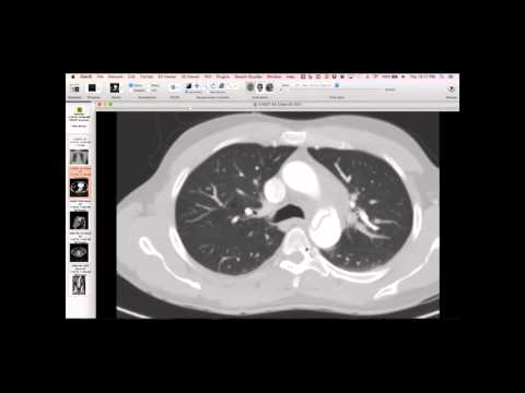
CT showed a markedly enlarged pulmonary artery. Lung parenchyma exhibited a mosaic attenuation pattern, patchy and extensive, with some opacity in the left lower lung. While no large filling defects were seen in the major vessels, there were dramatically abnormal vessels in the left lower lobe, appearing obliterated distally, and webs/stenosis in the right lower lobe vessels. Perfusion scans showed very abnormal perfusion, particularly in the left lower lobe. There was volume loss and apparent scarring in the left lower lobe, with scrawny pulmonary veins.
Key takeaways
This is a typical case of CTEPH, characterized by chronic pulmonary artery obstruction, pulmonary hypertension, and mosaic lung perfusion. The surgical report of pulmonary endarterectomy highlighted the difficulty in finding a dissection plane, especially in the more obliterated left lower lobe vessels, matching the imaging findings. The patient's left lower lobe demonstrated systemic arterial supply from transpleural collaterals rather than large bronchial arteries, potentially leading to edema. Slow blood flow and delayed contrast opacification in the pulmonary veins were also observed due to the severe vascular obstruction and collateral circulation.
AI-assisted summary — may contain errors. Verify against the source video. Learn more
More from this webinar
- Fibrovascular Polyp of the EsophagusNeoplastic
- Ectopic Thymoma in the Lower NeckNeoplastic
- Pentalogy of CantrellCongenital
- Acute Type B Aortic Dissection with Unilateral Pulmonary Artery HematomaVascular
- Rapidly Progressive Interstitial Lung Disease in Amyopathic DermatomyositisILD
- Lymphangioleiomyomatosis (LAM) Mimicking Alpha-1 Antitrypsin DeficiencyILD
- Acute Severe Pulmonary Embolism Requiring Surgical EmbolectomyVascular
- Pulmonary Artery Pseudoaneurysm in Cystic FibrosisVascular
See all cases from January 14, 2016 →