 CTICases
Granulomatosis with Polyangiitis
 ▶ Watch on YouTube — age-restricted, cannot embed here
[...]. Granulomatosis with Polyangiitis. CTICases. Published July 17, 2014. https://cticases.com/c/tkufx
Imaging findings
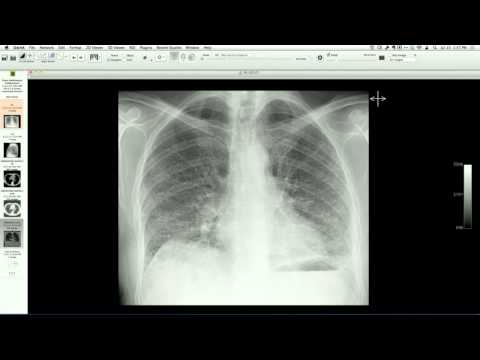
Chest CT demonstrates extensive, bilateral, poorly-marginated peribronchovascular consolidations and masses, with multiple areas of internal cavitation and associated diffuse alveolar hemorrhage. Serum proteinase 3 (PR3) levels are significantly elevated.
Key takeaways
Granulomatosis with polyangiitis (GPA) can occasionally present in an ICU setting with a highly aggressive, necrotizing vasculitis pattern. While multiple nodules or masses are classic, a dense peribronchovascular consolidation pattern with rapid cavitation and diffuse alveolar hemorrhage is a rare but well-described presentation of severe active disease that requires urgent immunosuppressive therapy.
AI-assisted summary — may contain errors. Verify against the source video. Learn more
More from this webinar
- Aortic Arch DissectionVascular
- Sclerosing PeriaortitisInflammatory
- Hypoplastic Left Heart Syndrome (Post-Norwood and Sano Shunt)Congenital
- Tetralogy of Fallot with Ductal Origin of the Left Pulmonary ArteryCongenital
- Migrating Pleural Mesothelial CystCongenital
- Chronic Diaphragmatic RuptureTrauma
- Bronchopulmonary DysplasiaCongenital
- Granulomatous-Lymphocytic Interstitial Lung DiseaseILD
See all cases from July 17, 2014 →