 CTICases
Asbestos-Related Pleural Plaques with Lung Distortion (Cheeto/Dorito Sign)
 ▶ Watch on YouTube — age-restricted, cannot embed here
[...]. Asbestos-Related Pleural Plaques with Lung Distortion (Cheeto/Dorito Sign). CTICases. Published January 14, 2016. https://cticases.com/c/v73qk
Imaging findings
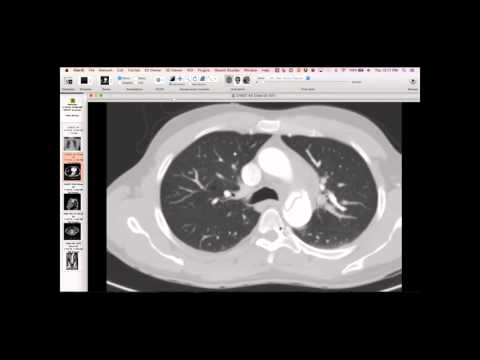
The patient presents with very large, exuberant pleural plaques, described as 'cheeto' and 'dorito' signs due to their thick, irregular, and sometimes tumefactive appearance. Around several plaques, there are gas pockets appearing next to the plaque, possibly representing torn lung or paracystic emphysema. There is also associated traction bronchiectasis, but some areas of gas don't appear like typical honeycombing, with relatively normal surrounding lung.
Key takeaways
These are dramatic asbestos-related pleural plaques, possibly causing mechanical distortion of the adjacent lung. The gas pockets might be due to the lung tearing or separating from adjacent alveolar walls as it attempts to move around the large, stiff plaques, forming small air pockets or paracystic emphysema. While indentation atelectasis and ground glass can be seen near plaques, this case also suggests true lung fibrosis with traction bronchiectasis and mechanical lung injury adjacent to the plaques, rather than purely asbestosis-related UIP.
AI-assisted summary — may contain errors. Verify against the source video. Learn more
More from this webinar
- Fibrovascular Polyp of the EsophagusNeoplastic
- Ectopic Thymoma in the Lower NeckNeoplastic
- Pentalogy of CantrellCongenital
- Acute Type B Aortic Dissection with Unilateral Pulmonary Artery HematomaVascular
- Rapidly Progressive Interstitial Lung Disease in Amyopathic DermatomyositisILD
- Lymphangioleiomyomatosis (LAM) Mimicking Alpha-1 Antitrypsin DeficiencyILD
- Acute Severe Pulmonary Embolism Requiring Surgical EmbolectomyVascular
- Pulmonary Artery Pseudoaneurysm in Cystic FibrosisVascular
See all cases from January 14, 2016 →
Related Exposure cases
- Silicone Injections and Pneumonitis
- Siderosis (Iron Deposition) from Occupational Exposure
- Necrotizing Fasciitis / Osteomyelitis from Heroin Skin Popping
- Pulmonary Langerhans Cell Histiocytosis (PLCH) - Classic Presentation
- Pulmonary Langerhans Cell Histiocytosis (PLCH) - Milder Spectrum (RBILD Transition)
- Bronchial Anthracofibrosis