 CTICases
Slow-Growing Low-Grade Chondrosarcoma of the Chest Wall
 ▶ Watch on YouTube — age-restricted, cannot embed here
[...]. Slow-Growing Low-Grade Chondrosarcoma of the Chest Wall. CTICases. Published January 14, 2016. https://cticases.com/c/2pz2u
Imaging findings
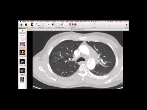
Serial imaging over three years shows a progressively enlarging, smooth-margined lesion arising in the chest wall, initially homogeneous with no matrix. Over time, it developed more dense areas and high-attenuation material, suggestive of calcification or blood. It was lifting the pectoralis muscle. Despite its growth, it lacked the destructive appearance or typical chondroid matrix expected of a high-grade chondrosarcoma. A right pleural effusion and pericardial effusion were noted on the most recent study.
Key takeaways
This case illustrates a very slow-growing, low-grade chondrosarcoma of the chest wall, which is unusual given its lack of prominent matrix on imaging and biopsy. Its slow growth rate over several years is also atypical for a chondrosarcoma, which usually progresses faster than a neurofibroma or schwannoma. The biopsy confirmed malignant chondrocytes without abundant matrix. The lesion's presentation prompted consideration of various diagnoses including neurofibroma or schwannoma due to its homogeneous appearance initially. Despite its low grade, it continued to grow and warranted resection.
AI-assisted summary — may contain errors. Verify against the source video. Learn more
More from this webinar
- Fibrovascular Polyp of the EsophagusNeoplastic
- Ectopic Thymoma in the Lower NeckNeoplastic
- Pentalogy of CantrellCongenital
- Acute Type B Aortic Dissection with Unilateral Pulmonary Artery HematomaVascular
- Rapidly Progressive Interstitial Lung Disease in Amyopathic DermatomyositisILD
- Lymphangioleiomyomatosis (LAM) Mimicking Alpha-1 Antitrypsin DeficiencyILD
- Acute Severe Pulmonary Embolism Requiring Surgical EmbolectomyVascular
- Pulmonary Artery Pseudoaneurysm in Cystic FibrosisVascular
See all cases from January 14, 2016 →