 CTICases
Mature Cystic Teratoma (Anterior Mediastinum)
 ▶ Watch on YouTube — age-restricted, cannot embed here
[...]. Mature Cystic Teratoma (Anterior Mediastinum). CTICases. Published July 17, 2014. https://cticases.com/c/df6bw
Imaging findings
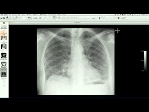
Chest radiograph shows an anterior mediastinal mass demonstrating the "hilum overlay sign," with a calcified rim. Contrast-enhanced chest CT confirms a well-circumscribed, rim-calcified anterior mediastinal mass containing macroscopic fat, low-attenuation fluid (sebaceous material), and soft-tissue elements.
Key takeaways
Mature cystic teratoma is the most common benign germ cell tumor of the mediastinum, typically occurring in the anterior mediastinum. While most frequently diagnosed in adolescents and young adults, they can be discovered incidentally in older adults and are characterized on CT by a mixture of fat, fluid, soft tissue, and calcification (often in the wall).
AI-assisted summary — may contain errors. Verify against the source video. Learn more
More from this webinar
- Aortic Arch DissectionVascular
- Sclerosing PeriaortitisInflammatory
- Hypoplastic Left Heart Syndrome (Post-Norwood and Sano Shunt)Congenital
- Tetralogy of Fallot with Ductal Origin of the Left Pulmonary ArteryCongenital
- Migrating Pleural Mesothelial CystCongenital
- Chronic Diaphragmatic RuptureTrauma
- Bronchopulmonary DysplasiaCongenital
- Granulomatous-Lymphocytic Interstitial Lung DiseaseILD
See all cases from July 17, 2014 →