 CTICases
Common Variable Immunodeficiency with Lymphocytic Interstitial Pneumonia
 ▶ Watch on YouTube — age-restricted, cannot embed here
[...]. Common Variable Immunodeficiency with Lymphocytic Interstitial Pneumonia. CTICases. Published October 30, 2015. https://cticases.com/c/bm8nc
Imaging findings
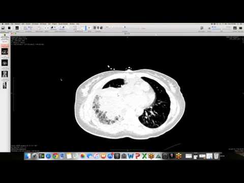
Serial CT studies show multiple bilateral nodular opacities with poorly defined margins and a mixture of ground-glass and solid attenuation, associated with cystic spaces and mild mediastinal lymphadenopathy. Over time, these opacities demonstrate a classic spontaneous "waxing and waning" pattern, with some nodules resolving and new ones developing without active treatment. PET CT shows that some of these lesions are FDG-avid.
Key takeaways
Common variable immunodeficiency (CVID) is frequently associated with non-infectious, benign lymphoproliferative disorders of the lung, such as lymphocytic interstitial pneumonia (LIP), follicular bronchiolitis, or granulomatous-lymphocytic interstitial lung disease (GLILD). A hallmark clinical and radiologic feature of these CVID-associated lymphoproliferative processes is their spontaneous waxing and waning appearance over time in the absence of therapy.
AI-assisted summary — may contain errors. Verify against the source video. Learn more
More from this webinar
- Beta-Thalassemia with Extramedullary Hematopoiesis and Diffuse Marrow ExpansionMetabolic
- Anomalous High Origin of the Right Coronary Artery with Incidental Developing PneumoniaCongenital
- Pulmonary Hemorrhage Secondary to Acute Pulmonary Embolism presenting as Abdominal PainVascular
- Acute Exacerbation of Idiopathic Pulmonary Fibrosis Triggered by Minor TraumaILD
- Type A Intramural Hematoma with Focal Intimal TearVascular
- Aortic Rupture and Mediastinal Hematoma Decompressing into the Right LungVascular
- Congenital Left Main Coronary Artery Osteal AtresiaCongenital
- Pleural Extramedullary Hematopoiesis in MyelofibrosisMetabolic
See all cases from October 30, 2015 →
Related ILD cases
- Desquamative Interstitial Pneumonia (DIP) in a Heavy Smoker
- Anti-Synthetase Syndrome (Anti-Jo-1) with Organizing Pneumonia
- Fibrotic Organizing Pneumonia
- Restrictive Allograft Syndrome (Post-Transplant Upper Lobe Progressive Fibrosis)
- Bleomycin-Induced Severe Alveolar Damage with Barotrauma
- Hypersensitivity Pneumonitis (HP) in a Dog Groomer