 CTICases
Central Bronchial Carcinoid Tumor
 ▶ Watch on YouTube — age-restricted, cannot embed here
[...]. Central Bronchial Carcinoid Tumor. CTICases. Published August 20, 2015. https://cticases.com/c/mn4wy
Imaging findings
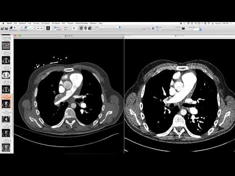
Chest radiographs showed large, rounded, non-segmental opacities in the lower lung zones, with unilateral pulmonary artery enlargement on frontal view, and a dramatic lesion on lateral view. CT showed a central bronchial mass, located in the superior segment of the left lower lobe, with dramatic calcifications internally.
Key takeaways
A central bronchial mass with calcifications, especially when presenting with unilateral pulmonary artery enlargement or a dramatic radiographic opacity, is highly suggestive of a bronchial carcinoid tumor. The combination of these findings virtually eliminates other differential diagnoses.
AI-assisted summary — may contain errors. Verify against the source video. Learn more
More from this webinar
- Retained Wires from First-Generation DefibrillatorIatrogenic
- Hyperacute Pulmonary Infarction progressing to Acute PE with Pleural EffusionVascular
- Left Upper Lobe Anomalous Pulmonary Venous Stricture (Incidental)Congenital
- Lymphocytic Interstitial Pneumonia (LIP) in association with B-cell LymphomaNeoplastic
- Endobronchial Papillomatosis with Left Lower Lobe CollapseNeoplastic
- Esophageal Carcinoma with Radiographic CorrelationNeoplastic
- Cardiogenic Pulmonary Edema with Ground Glass OpacityVascular
- Hypersensitivity Pneumonitis (HP) in a Dog GroomerILD
See all cases from August 20, 2015 →
Related Neoplastic cases
- Choriocarcinoma with Hemorrhagic Pulmonary Metastases
- Adenocarcinoma Spectrum (Preinvasive and Invasive Components)
- Pericardial Vascular Malformation (Veno-Hemangioma with Phleboliths)
- Recurrent Squamous Cell Carcinoma in Respiratory Papillomatosis with Rib Invasion
- Ectopic Mediastinal Parathyroid Adenoma
- Post-Transplant Lymphoproliferative Disorder (PTLD) Presenting as Airway-Centric Lung Nodules