 CTICases
Intracardiac Mature Cystic Teratoma
 ▶ Watch on YouTube — age-restricted, cannot embed here
[...]. Intracardiac Mature Cystic Teratoma. CTICases. Published July 17, 2014. https://cticases.com/c/rkq5a
Imaging findings
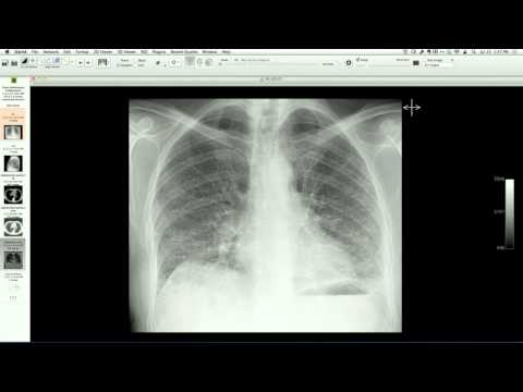
Cardiac MRI demonstrates a large, heterogeneous, lobulated mass within the right ventricle, showing high signal intensity on T2-weighted and black-blood sequences with foci of fat and proteinaceous fluid. The mass is adherent to the tricuspid valve apparatus and papillary muscles, and pathology post-resection confirms a mature cystic teratoma containing multiple tissue types, including cystic spaces and focal brain tissue.
Key takeaways
Intracardiac teratomas are exceedingly rare benign germ cell tumors, typically diagnosed in infants and children but occasionally presenting in adults. They most commonly arise within the pericardial space, and primary intracardiac (cavity) presentation is highly unusual, requiring surgical resection and potentially valve replacement if the tumor is densely adherent to the subvalvular apparatus.
AI-assisted summary — may contain errors. Verify against the source video. Learn more
More from this webinar
- Aortic Arch DissectionVascular
- Sclerosing PeriaortitisInflammatory
- Hypoplastic Left Heart Syndrome (Post-Norwood and Sano Shunt)Congenital
- Tetralogy of Fallot with Ductal Origin of the Left Pulmonary ArteryCongenital
- Migrating Pleural Mesothelial CystCongenital
- Chronic Diaphragmatic RuptureTrauma
- Bronchopulmonary DysplasiaCongenital
- Granulomatous-Lymphocytic Interstitial Lung DiseaseILD
See all cases from July 17, 2014 →